Brain Anatomy in Treatment-Resistant Schizophrenia: A Signal, Not a Test
A large international imaging study found group-level structural differences in treatment-resistant schizophrenia, but the measurements cannot yet identify patients individually or replace clinical judgment.

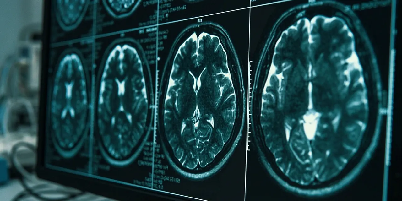
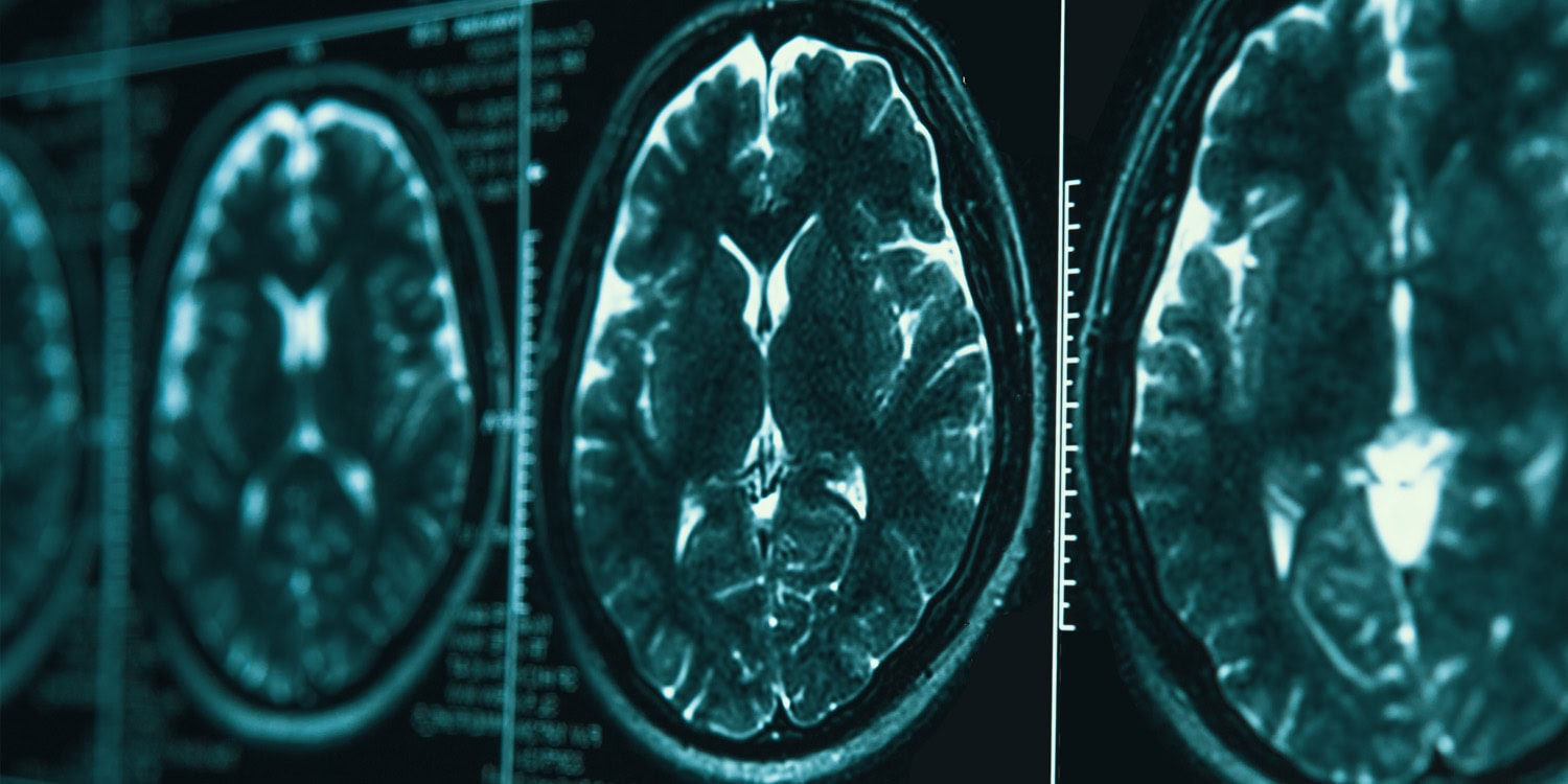
A multi-site imaging study in Molecular Psychiatry reports that people whose schizophrenia has not yielded to usual antipsychotics differ in brain anatomy from those who respond to treatment. The work was headed by Semra Etyemez, Kun Yang and Akira Sawa of Johns Hopkins University School of Medicine. Compared with responsive patients, the resistant group had lower tissue volume on each side of the hippocampus, a seahorse-shaped structure tied to memory and learning. The study also found less putamen volume and a smaller surface area in the left superior frontal gyrus. The authors present these as group-level differences that might someday help flag likely nonresponders. [1]
The regional pattern was not uniform. In the hippocampus and the superior frontal gyrus, the data followed a graded ordering: healthy participants had the most tissue, responsive patients less, and resistant patients least. The putamen broke that pattern. Resistant patients did not differ statistically from healthy controls there, while responsive patients had a larger putamen than healthy controls. That inconsistency is part of the result, not a footnote. It cautions against treating all three regions as equivalent signals and leaves the putamen's role unresolved in the source. [1]
The authors frame the results as a step toward earlier alternative interventions. The report says the variations could eventually help doctors identify patients unlikely to benefit from typical antipsychotics. That is a forecast about possible clinical use, not an outcome demonstrated in the paper. The same report notes that the measurements cannot yet diagnose resistance on their own. The word eventually matters: it separates a promising group-level signal from a tool that clinicians could use today. [1]
The investigators worked with an international neuroimaging consortium and analyzed scans from over 1,200 people at seven sites in six countries. The sample comprised 158 patients with treatment-resistant schizophrenia, 599 patients who responded to standard drugs, and 508 healthy comparison participants. Clinicians at each site sorted patients using clinical and medication records. The sites spanned North America, Europe and Asia. This design gave the researchers a larger base than earlier smaller studies, but the source does not describe the sample as a population-representative survey. [1]
Advertisement
Two analytic routes were used. First, a meta-analysis combined results from independent datasets to look for broad trends in deep-brain volumes and in the thickness and surface area of the cortex. Models were adjusted for age, sex, race, head size and duration of illness. Second, a mega-analysis pooled raw scan data into one dataset and added healthy participants as a baseline. The second pass reproduced the hippocampal, putamen and left frontal findings. Agreement across methods strengthens the group-level observation; it does not turn a scan into an individual diagnosis. [1]
The clinical stakes are high because treatment resistance is common and hard to identify early. Most standard antipsychotics work by blocking dopamine receptors, and many patients improve on them. Yet the report states that as many as 60 percent do not get adequate relief from such drugs. Clozapine is the sole medication approved for this group, and it requires regular blood checks because of dangerous immune-related complications. Even with clozapine, a substantial share of patients do not respond. The source therefore frames a biological marker as a route to earlier alternatives, not as a replacement for careful treatment trials. [1]
At present, clinicians label treatment resistance only after multiple antipsychotic trials fail. That process can stretch across months or years, and the report notes that mental health often declines during the wait. An objective marker could, in principle, shorten that interval and prompt specialized care sooner. The new study matters because it identifies structural differences that might contribute to such a marker. It does not show that the marker exists in usable form. The authors describe the anatomy as a possible component of future risk assessment, not as a current standard. [1]
Earlier, smaller imaging projects had pointed toward anatomical shifts in treatment-resistant schizophrenia, but the researchers sought a wider dataset to test whether those patterns held. The new work is best understood as a large-scale check on prior hints. The condition itself involves hallucinations, delusions and cognitive problems. Standard drugs reduce symptoms for many by blocking dopamine receptors, but the report says treatment resistance remains a major challenge in psychiatric care. That background explains why a structural finding attracts interest even when it cannot yet guide practice. [1]
Advertisement
Medication exposure varied in type, amount and duration, so the team examined whether drug history explained the anatomy. Cumulative antipsychotic dosage was not statistically significant for hippocampal size or for the surface area of the superior frontal gyrus. The researchers also checked clozapine use. That analysis supports, but does not prove, the interpretation that those two differences track the underlying biology of treatment resistance rather than the drugs themselves. The source's wording is cautious: it says the lack of association suggests such a link, not that it establishes one. [1]
The putamen behaved differently. Its volume had a weak correlation with cumulative medication use, raising the possibility that antipsychotics slightly affect this structure. The group comparison adds another wrinkle: resistant patients and healthy controls did not differ statistically in putamen size, while responsive patients had a larger putamen than healthy controls. The source does not explain why. For readers, the practical point is that the three regions do not tell one simple story. The hippocampal and frontal results are cleaner than the putamen result, and the authors do not claim otherwise. [1]
The medication analysis has boundaries of its own. It relates cumulative dosage to structure, but the source does not describe a controlled experiment or a full causal model. The finding for the hippocampus and frontal surface is framed as a lack of a statistically significant dosage association, not as proof that drugs play no role. For the putamen, the weak correlation leaves room for medication influence. These distinctions matter because treatment-resistant patients often have long and complex drug histories, and the source does not describe the study as a controlled trial. [1]
The sample has a demographic gap. Although data came from six countries in North America, Europe and Asia, the pool lacked African and Latino representation. The research team said future neuroimaging should include more diverse demographics to test whether the anatomical differences hold across all human populations. That is a question about generalizability. It does not erase the current findings, but it limits how far they can be extended before broader samples are examined. [1]
Advertisement
The comparison group carries its own uncertainty. Patients classified as responsive might include people who will develop treatment resistance later. The source raises that possibility but does not quantify it. If such a shift occurs, the contrast between groups could be blurred in ways the study cannot measure. The report also does not establish whether the observed anatomical differences come before, alongside or after the emergence of resistance. Those are open questions, not conclusions. The current design can show association at a point in time; it cannot settle timing or causation. [1]
Most important for clinical use, the brain measurements failed an individual-level test. When the researchers checked predictive power, the models could not reliably separate treatment-resistant patients from other schizophrenia patients one person at a time. A statistically robust average difference can still be a poor diagnostic tool. The source says the structural differences are not yet enough on their own to diagnose resistance. It points toward joining imaging with other biological indicators, such as genetic profiles or metabolic markers, to build a reliable predictive test. That combination is a research goal, not an available product. [1]
Several mechanistic questions remain. The report does not identify a biological pathway connecting the structural measurements to medication response. It does not say whether early intervention could alter the trajectory of these brain regions. It also does not establish that the hippocampal or frontal differences are causes rather than correlates. The medication checks reduce one alternative explanation for those two regions, but they do not eliminate every possible confound. These gaps are part of the study's limits, and the source does not present them as resolved. [1]
The next questions are specific. Will more diverse samples reproduce the hippocampal and left frontal pattern? Can imaging combined with genetic or metabolic data improve prediction beyond scans alone? Would any such model identify resistance early enough to change care without exposing patients to unnecessary alternatives? These are scenarios and questions, not forecasts of success. The report explicitly calls for broader demographic representation and for work that combines brain imaging with other biological indicators. It does not propose a timeline, a validated algorithm or a change to current practice. [1]
Advertisement
Another watch item is how any future marker would fit alongside clozapine safety rules. Clozapine remains the only approved drug for this population and still requires blood monitoring for severe immune risks. A future pathway would need to respect those constraints and would still face the fact that some patients do not respond to clozapine. For now, the study adds strong group-level evidence for structural differences, especially in the hippocampus and left superior frontal gyrus, while leaving the putamen and individual prediction unresolved. The clinical question remains open. [1]
Sources
Prepared with AI assistance from the cited sources.
The Cali Reporter newsletter
The stories worth your time.
California, the country, and what comes next. Join our free newsletter for reporting that keeps you informed.
More in Relationships
- Family Relationships May Influence Well-Being of Poor Taiwanese Adolescents
A study of Taiwanese adolescents in poverty suggests that family relationships may play a key role in explaining the link between material deprivation and psychological distress, as well as substance use.
- Aggressive Personality Traits and Jealousy in Romantic Relationships
A new study explores how aggressive personality traits and specific love styles contribute to jealousy in romantic relationships.
- Early Brain Differences in Dyslexia Linked to Visual Processing Before Formal Reading
A longitudinal study published in Developmental Cognitive Neuroscience reveals that children who later develop dyslexia already show distinct neural responses to written words in kindergarten, before formal reading…